
Echo viruses (Enteric, cytopatogenic Human Orphan viruses) are members of the genus Enterovirus of the Picornaviridae family. They constitute the largest subset of enteroviruses, consisting of 34 serotypes. Echo viruses cause a variety of diseases, from minor febrile conditions to severe, potentially fatal conditions such as childhood enteritis, aseptic meningitis, encephalitis, paralysis and myocarditis.
Initially, Echo viruses were classified into 34 serotypes. Echo-virus 10 is reclassified as reovirus 1 and Echo-virus 28 as rhinovirus 1. Echo-virus 9 is now recognized as the same as Coxisakivirus A23. Echo-virus 22 and 23 are reclassified as members of the genus Parechovirus, and Echo-virus 34 is a variant of coxakivirus A24.
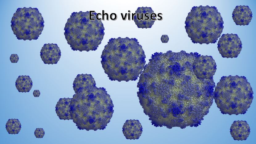
Structure and cultivation
The structure of ECHO viruses is similar to that of other enteroviruses – virus particles have ixahedral symmetry and are composed of RNA and proteins. Their capsid is composed of 60 subunits and is formed from 4 proteins – VP1 to VP4. These proteins play an important role in determining host range and tropism of the virus. Echoviruses are infectious in a wide range of pH 3 to 10 and are resistant to the effects of ether and alcohols. Monolayer cell cultures of human embryonic trophoblasts are most favorable for viral propagation. Your oral particles are not pathogenic to experimental animals except for some strains that adapt to newborn mice.
Antigenic structure
There are 34 known serotypes of Echo viruses, differing only in complement binding response and viral neutralization response. Some viral strains have hemagglutinins, which agglutinate human red blood cells.
Epidemiology
Resistance of Echo viruses to environmental factors is high, with external storage for months. They are sensitive to chlorine preparations such as chloramine and lime. The natural reservoir and source is man. The infection is spread by air-droplet and fecal-oral mechanism. More than 90% of echovirus infections are asymptomatic. The incidence of infections varies depending on the season, geographical location, and age of the population, and are more common in the summer and early fall.
People of all ages are vulnerable to infection with Echo Viruses. According to the World Health Organization, children under the age of 15 are most susceptible to infection. Outbreaks have been reported in Panama, Mexico, Switzerland, Cuba, the United States and Turkey.
Pathogenesis and clinical presentation
Viral replication of some strains takes place in the pharynx and then spreads to regional lymph nodes. Most viral particles in the faecal-oral mechanism reach the digestive system, where the virus binds to specific receptors. After replication, echo viruses spread to regional lymph nodes and cause subclinical transitional viremia. During this low-grade viremia, the virus spreads to the liver, spleen, bone marrow and distant lymph nodes. Secondary sites of infection include CNS, liver, spleen, bone marrow, heart and lungs.
Most often, ECHO viruses cause fever, respiratory and flu-like illness, aseptic meningitis, but 90% of patients with echoviral infection are asymptomatic. The most common presentation is the presence of fever. Infection can be caused by almost any enterovirus serotype and is clinically indistinguishable from infection with many other viral agents.
Echo-viral meningitis may have a two-phase pattern – fever and myalgia with clinical immobilization, followed by recurrence of fever and headache, mark the onset of aseptic meningitis.
Skin rashes are more common in echovirus infections than in other enterovirus infections. The exanthems can be maculopapulous, macular or papuopustular.
Microbiological diagnostics
Investigating:
- throat secretion;
- feces;
- blood;
- liquor;
- washes from the patient’s objects.
Pre-treatment with antibiotic agents of the collected samples is mandatory in order to eliminate the presence of bacterial microflora. During the first week of echoviral infection, the virus is detected in the larynx, and in the next 3 to 5 weeks it is detected by microbiological examination of faeces. Viral isolation is not accomplished by infecting cell cultures, and typing by a viral neutralizing reaction. Viral neutralizing antibodies appear within the first week of the disease, making it possible to apply serological diagnostics.
Diagnosis
Echo viruses are detected with a method, called PCR. There are available kits, both conventional and RT PCR that can detect the presence.